- Retrait gratuit dans votre magasin Club
- 7.000.000 titres dans notre catalogue
- Payer en toute sécurité
- Toujours un magasin près de chez vous
- Retrait gratuit dans votre magasin Club
- 7.000.000 titres dans notre catalogue
- Payer en toute sécurité
- Toujours un magasin près de chez vous
Description
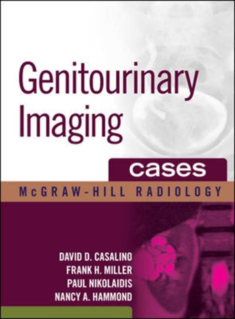
295 cases and more than 1700 illustrations teach you how to accurately interpret genitourinary tract images
4 STAR DOODY'S REVIEW!
"The high-quality images and pithy discussions make this book very useful to radiologists, both in training and in practice....The book's best features are the excellent image quality, the inclusion of images of differential diagnostic considerations, and concise discussions of the cases. This is an excellent resource for radiologists in training and in practice. The case-based format is excellent for board preparation, and its concise prose provides all the necessary information while leaving out the excess."--Doody's Review Service
Genitourinary Imaging Cases presents an efficient and systematic approach to examining images of the genitourinary system. You will find an unmatched collection of 295 cases ranging from normal anatomy to the full spectrum of disease -- including renal cystic masses, renal infection, renal vascular disease, and female pelvic abnormalities. Included with these cases are 1700+ high-quality images that are representative of what you would see on various imaging modalities.
The book's easy-to-navigate organization is specifically designed for use at the workstation. The concise text, numerous images, and helpful icons speed access to essential information and simplify the learning process.
Features:
- Each case includes findings, differential diagnosis, comment/discussion, and clinical pearls
- Icons, a grading system depicting the full spectrum of diseases, common to rare, and imaging findings, typical to unusual, along with the consistent chapter organization make this perfect for rapid at-the-bench consultation
- Strong focus on pathology
- Special emphasis on the latest diagnostic modalities that include both CT and MR images
Spécifications
Parties prenantes
- Auteur(s) :
- Editeur:
Contenu
- Nombre de pages :
- 672
- Langue:
- Anglais
- Collection :
Caractéristiques
- EAN:
- 9780071479127
- Date de parution :
- 05-02-10
- Format:
- Livre relié
- Format numérique:
- Genaaid
- Dimensions :
- 216 mm x 277 mm
- Poids :
- 2222 g


