- Retrait gratuit dans votre magasin Club
- 7.000.000 titres dans notre catalogue
- Payer en toute sécurité
- Toujours un magasin près de chez vous
- Retrait gratuit dans votre magasin Club
- 7.000.0000 titres dans notre catalogue
- Payer en toute sécurité
- Toujours un magasin près de chez vous
34,95 €
+ 69 points
Format
Description
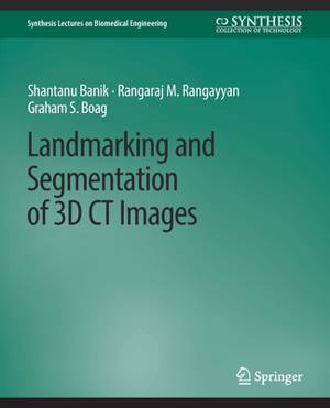
Segmentation and landmarking of computed tomographic (CT) images of pediatric patients are important and useful in computer-aided diagnosis (CAD), treatment planning, and objective analysis of normal as well as pathological regions. Identification and segmentation of organs and tissues in the presence of tumors are difficult. Automatic segmentation of the primary tumor mass in neuroblastoma could facilitate reproducible and objective analysis of the tumor's tissue composition, shape, and size. However, due to the heterogeneous tissue composition of the neuroblastic tumor, ranging from low-attenuation necrosis to high-attenuation calcification, segmentation of the tumor mass is a challenging problem. In this context, methods are described in this book for identification and segmentation of several abdominal and thoracic landmarks to assist in the segmentation of neuroblastic tumors in pediatric CT images. Methods to identify and segment automatically the peripheral artifacts and tissues, the rib structure, the vertebral column, the spinal canal, the diaphragm, and the pelvic surface are described. Techniques are also presented to evaluate quantitatively the results of segmentation of the vertebral column, the spinal canal, the diaphragm, and the pelvic girdle by comparing with the results of independent manual segmentation performed by a radiologist. The use of the landmarks and removal of several tissues and organs are shown to assist in limiting the scope of the tumor segmentation process to the abdomen, to lead to the reduction of the false-positive error, and to improve the result of segmentation of neuroblastic tumors. Table of Contents: Introduction to Medical Image Analysis / Image Segmentation / Experimental Design and Database / Ribs, Vertebral Column, and Spinal Canal / Delineation of the Diaphragm / Delineation of the Pelvic Girdle / Application of Landmarking / Concluding Remarks
Spécifications
Parties prenantes
- Auteur(s) :
- Editeur:
Contenu
- Nombre de pages :
- 148
- Langue:
- Anglais
- Collection :
Caractéristiques
- EAN:
- 9783031005077
- Date de parution :
- 18-03-09
- Format:
- Livre broché
- Format numérique:
- Trade paperback (VS)
- Dimensions :
- 190 mm x 235 mm
- Poids :
- 303 g

Les avis
Nous publions uniquement les avis qui respectent les conditions requises. Consultez nos conditions pour les avis.


