- Retrait gratuit dans votre magasin Club
- 7.000.000 titres dans notre catalogue
- Payer en toute sécurité
- Toujours un magasin près de chez vous
- Retrait gratuit dans votre magasin Club
- 7.000.0000 titres dans notre catalogue
- Payer en toute sécurité
- Toujours un magasin près de chez vous
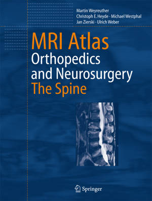
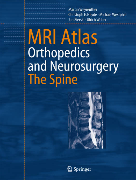
MRI Atlas
Orthopedics and Neurosurgery, the Spine
Martin Weyreuther, Christoph E Heyde, Michael Westphal, Jan Zierski, Ulrich WeberDescription
This atlas of spinal MRI is the fruit of interdisciplinary cooperation among radiologists, orthopedic surgeons, traumatologists, and neurosurgeons. It is clinically oriented, covering all important diseases and injuries of the spine. Numerous illustrations are supplemented by concise descriptions of anatomy and pathophysiology, normal and abnormal MRI appearance, diagnostic pitfalls, and the clinical significance of MRI. The contents are arranged in a logical, accessible sequence, for quick reference: Normal Anatomy and Variants; Congenital and Developmental Anomalies; Trauma and Fractures; Degenerative Changes; Inflammatory Conditions; Tumors and Tumor-like Lesions; Postoperative Changes; and Suggested Reading. The clear and didactic style enables readers to assimilate the fundamentals of spinal anatomy and disease states as a basis for understanding diagnostic strategies and surgical management. By combining descriptions of the clinical manifestation of spinal disorders with the corresponding MRI findings, the book helps readers to develop a meaningful approach to the interpretation of MRI of the spine.
Spécifications
Parties prenantes
- Auteur(s) :
- Traducteur(s):
- Editeur:
Contenu
- Nombre de pages :
- 298
- Langue:
- Anglais
Caractéristiques
- EAN:
- 9783540335337
- Date de parution :
- 13-10-06
- Format:
- Livre relié
- Format numérique:
- Genaaid
- Dimensions :
- 281 mm x 215 mm
- Poids :
- 1097 g

Les avis
Nous publions uniquement les avis qui respectent les conditions requises. Consultez nos conditions pour les avis.


