- Retrait gratuit dans votre magasin Club
- 7.000.000 titres dans notre catalogue
- Payer en toute sécurité
- Toujours un magasin près de chez vous
- Retrait gratuit dans votre magasin Club
- 7.000.0000 titres dans notre catalogue
- Payer en toute sécurité
- Toujours un magasin près de chez vous
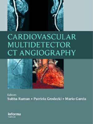
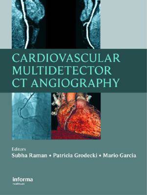
Cardiovascular Multidetector CT Angiography
Subha Raman, Patricia Grodecki, Mario GarciaDescription
Cardiovascular medicine has witnessed significant progress over the past century, incorporating the technical advances of each era to improve patient care. The introduction of the stethoscope, electrocardiography, roentgenography, angiography, invasive hemodynamics, ultrasonography, nuclear scintigraphy, and magnetic resonance have each, in turn, allowed progressively greater accuracy and precision in the diagnosis and treatment of cardiovascular disease.
The advent of multidetector computed tomography (MDCT) using 64 detector rows provides the next leap forward in cardiovascular care by delivering on the promise of high-resolution visualization of cardiovascular structure and function noninvasively.
Cardiovascular Multidetector CT Angiography demonstrates the clinical context within which this technology is useful for individual patient assessment, providing the technical information needed to perform cardiovascular CTA and focusing on the spectrum of clinical applications.
Written by acknowledged experts in the CTA arena, the book contains an abundance of 64-slice images--the highest technical resolution available--to identify disease states. Topics covered include normal cardiac anatomy, abnormal coronary arteries, coronary anomalies, artifacts, left and right ventricular function and abnormalities, valvular heart disease, pericardium, and the aorta. The book also discusses cardiac masses, cardiac veins, peripheral artery disease, and congenital heart disease.
Demonstrating the application of cardiovascular multidetector computed tomography from the perspective of patient care, the book is composed entirely of case studies--an excellent format for teaching those just beginning to work with CTA.
The book is designed for cardiologists and radiologists alike, as well as primary care physicians, medical students, and other health care professionals who have the opportunity to use this technology to improve diagnosis and treatment for their patients with cardiovascular disease.
Spécifications
Parties prenantes
- Auteur(s) :
- Editeur:
Contenu
- Nombre de pages :
- 190
- Langue:
- Anglais
Caractéristiques
- EAN:
- 9781841846453
- Date de parution :
- 30-07-07
- Format:
- Livre relié
- Format numérique:
- Genaaid
- Dimensions :
- 222 mm x 290 mm
- Poids :
- 1056 g

Les avis
Nous publions uniquement les avis qui respectent les conditions requises. Consultez nos conditions pour les avis.


