- Retrait gratuit dans votre magasin Club
- 7.000.000 titres dans notre catalogue
- Payer en toute sécurité
- Toujours un magasin près de chez vous
- Retrait gratuit dans votre magasin Club
- 7.000.0000 titres dans notre catalogue
- Payer en toute sécurité
- Toujours un magasin près de chez vous
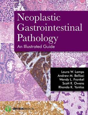
Neoplastic Gastrointestinal Pathology: An Illustrated Guide
An Illustrated Guide
Laura Lamps, Andrew Bellizzi, Wendy L Frankel, Scott R Owens, Rhonda YantissDescription
"This book is very easy to read, well organised, beautifully illustrated and full of quick-reference tables. The authors also provide practical advice on dealing with controversial and difficult areas... a welcome new addition for the pathology reporting room. Trainee pathologists will find the 'general approach' chapters very helpful and educational. Practising pathologists will be able to access the quick-reference tables and numerous illustrations in their day-to-day practice."
--Professor Fiona Campbell, Histopathology, Royal Liverpool and Broadgreen, University Hospitals, Gut
This concisely written, abundantly illustrated guide to a wide range of topics in gastrointestinal neoplasia facilitates the evaluation and accurate diagnosis of gastrointestinal neoplasms, both straightforward and challenging. This approachable guide covers the entire tubular GI tract and features more than 600 high quality images.
Written and arranged with the busy practicing pathologist in mind, this practical guide includes, for each entity, definitions and terminology, gross and morphologic features, differential diagnoses, useful ancillary tests, staging and grading parameters, and clinical considerations. Beautiful color figures throughout the volume highlight essential histologic features as well as differential diagnoses and potential diagnostic pitfalls. The book is organized into six introductory chapters focused on approaches to neoplasia, followed by 6 organ-specific chapters covering each segment of the GI tract. The final two chapters offer an in-depth discussion of immunohistochemistry and molecular pathology of gastrointestinal neoplasia. This book is a vital reference for practicing pathologists, and with its clear, concise presentation it is also an excellent resource for pathologists in training.
Key Features:
- Provides clear, concise coverage of neoplastic disease across the entire tubular gastrointestinal tract
- Offers over 600 high-quality images highlighting key differential points and potentially misleading variants
- Comprehensive tables cover diagnostic features, tumor types, and other crucial data for pathologists
Spécifications
Parties prenantes
- Auteur(s) :
- Editeur:
Contenu
- Nombre de pages :
- 408
- Langue:
- Anglais
Caractéristiques
- EAN:
- 9781936287727
- Date de parution :
- 15-07-15
- Format:
- Livre relié
- Format numérique:
- Genaaid
- Dimensions :
- 221 mm x 287 mm
- Poids :
- 1542 g

Les avis
Nous publions uniquement les avis qui respectent les conditions requises. Consultez nos conditions pour les avis.


